As you may have noticed, there’s a lot less stuff going up here as I’m full-time teaching anatomy to undergrads these days. This is a lot of fun and a bit of a challenge, trying to communicate anatomical and clinical concepts to a bunch of 18 year olds who aren’t sure of their lateral from their medial yet.
I have a whole bunch of lectures prepared and given but I want to try and find some way of getting all the work I put into them online.
We have a great collection of anatomical images that we can use in the department, but there are copyright restrictions involved and confidentiality issues involved in the use of images of our donor bodies, so I’m gonna try and cover some of the key/interesting parts of the lectures using whatever copyright free images I can find.
They are of course going to be more student orientated than fellowship orientated but there still might be something you can learn. Let me know if you find them useful or awful…
The Cervical Spine – Some things worth knowing
You have 7 cervical vertebra and 8 paired cervical spinal nerves. Why?
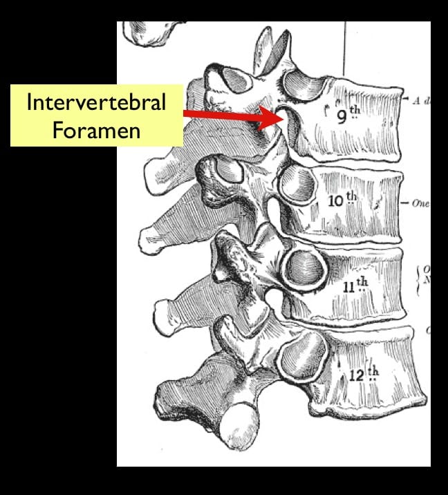
- normally the cervical spinal nerves are named for the vertebra that they emerge under; T9 spinal nerve emerges in the intervertebral foramen between T9 and T10
- the first cervical spinal nerve (C1/sub-occipital nerve) emerges above C1 vertebra (actually between the skull and C1)
- therefore C7 spinal nerve emerges above the C7 vertebra and the C8 spinal neve emerges below the C7 vertebra
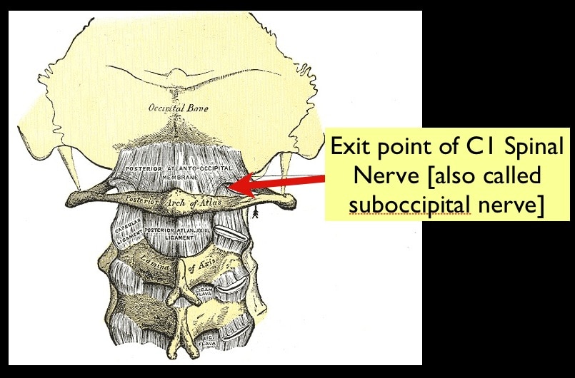
Worth noting that the greater occipital nerve that is blocked in the treatment of headache is actually the post rami of C2. Not the sub-occipital nerve.
This guy has a unique technique of treating headaches using para-spinal injection of bupivicaine and has great results. But I digress…
What is the course of the vertebral artery?
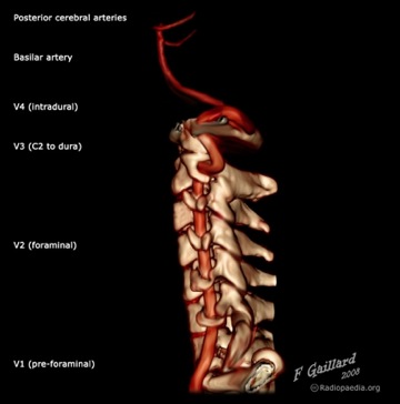
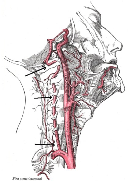
- the vertebral artery as its origin from the 1st part of the subclavian
- passes posteriorly and superiorly to enter the transverse foramen (only cervical vertebra have these) of C6 (not C7!)
- it ascends vertically in the foramen with a sympathetic nervous plexus and a vertebral vein
- after emerging from C1 it makes a 90 degree turn posterior and medially along the post arch of the atlas
- it then turns again and pierces the posterior atlanto-occiptal membrane before passing through the foramen magnum into the skull
- The eccentric course is thought to be responsible for dissections that occur following blunt trauma.
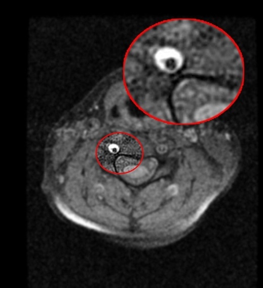
- Also worth noting that the left is normally substantially bigger than the right.
Some Ligaments of the C-spine:
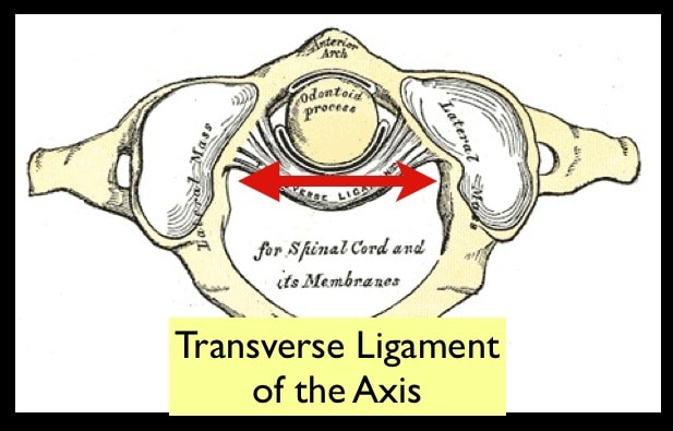
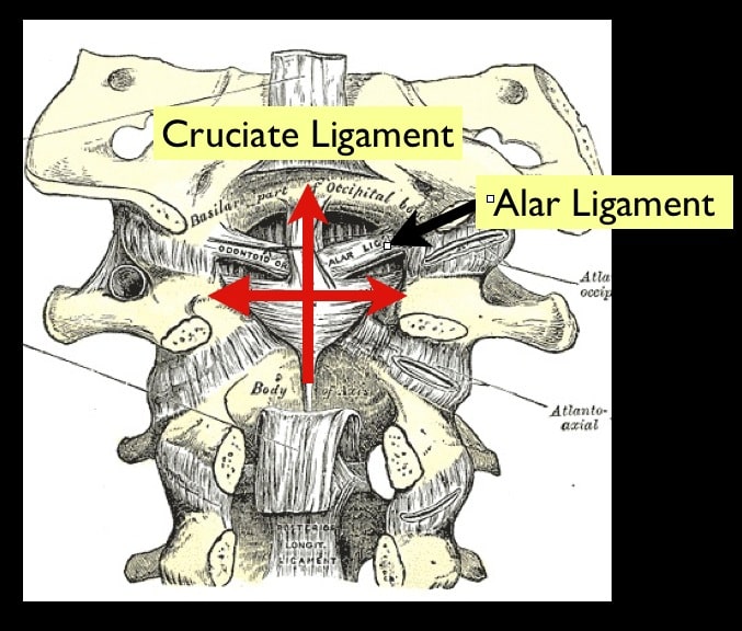
- The Ligaments connecting the occiput, atlas and axis together are the most important from our point of view
- in particular the transverse ligament is key in holding the atlas firmly against the dens of the axis.
- If the ligament ruptures you can problems like this (16 secs to 22 secs the best bit)
- or the ligament may break the dens right off the axis like this:
- Or if you like a static image:
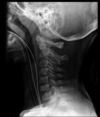
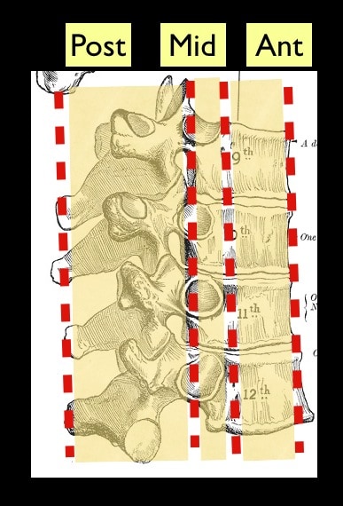
What about the columns?
- the spine is commonly split up into three columns (by this guy PMID 6670016)
- Anterior – ant longitudinal ligament and ant 2/3 of vertebral bodies
- Middle – post longitudinal ligament and post 1/3 of vertebral bodies
- Posterior – ligamentum flavum and pedicles, lamina, spinous processes
- An injury that covers more than 1 column is more likely to be unstable
- There are considered to be multiple fascial compartments or spaces in the head and neck. The main fascial compartments in the neck are:
- the carotid sheaths
- the prevertebral fascia
- the pretracheal fascia

- Pus can spread along these compartments from the pharyngeal and sub-mandibular and sub-lingual spaces to the mediastinum

Excellent post, thanks for this. Any more?
Thanks for the feedback
I’m working on it!
I feel quite nauseated Andy – Great videos!
Most excellent post and I am always continuing to learn!
cheers for the kind comments!
Absolute love your podcasts! Please can you do some more
Thanks sonya
I have a whole bunch planned but the small children are kind of holding me back from getting them finalised