[This is a series of posts where I’m trying to transfer my anatomy lectures to a blog format, with emphasis on the clinically relevant points.
Images are mainly from the 1918 Grays, freely available and out of copyright here. Forgive the out of date nomenclature…]
The Nasal Cavity
- the nasal cavity is a large air filled space at the centre of the face
- it is surrounded by other air-filled spaces
I know that much is obvious but it’s important for understanding how when one space goes wrong, often another one does too.
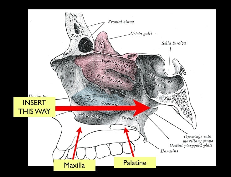
The floor of the nasal cavity is the roof of the oral cavity and made up of maxilla and palatine bone.
- Everybody has watched someone trying to put an NP airway almost vertically up the nose, instead of along the floor of the nasal cavity. This applies to any tubes you’re sticking in the nose.
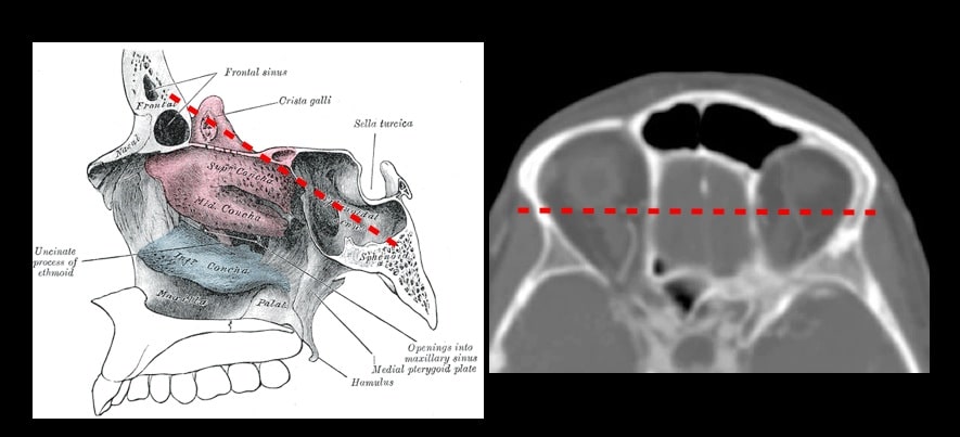
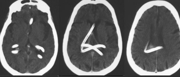
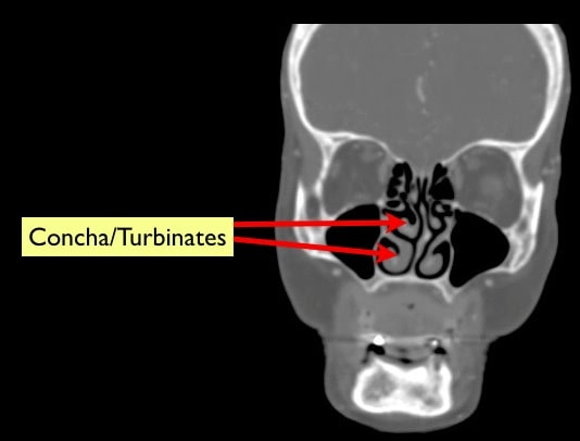
- fractures through the cranial vault as shown above can allow 2 way passage
- CSF out
- medical interventions in (see CT below)
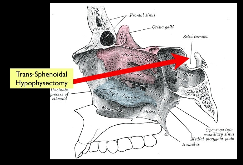
- one exceptionally cool thing the nasal cavity is utilised for is for pituitary adenoma removal
- If you have 6 mins to spare and you’re interested the video below is fascinating to see it in action
Epistaxis
- Anteriorly we’re talking Little’s area, the anastamotic point on the cartilagenous septum
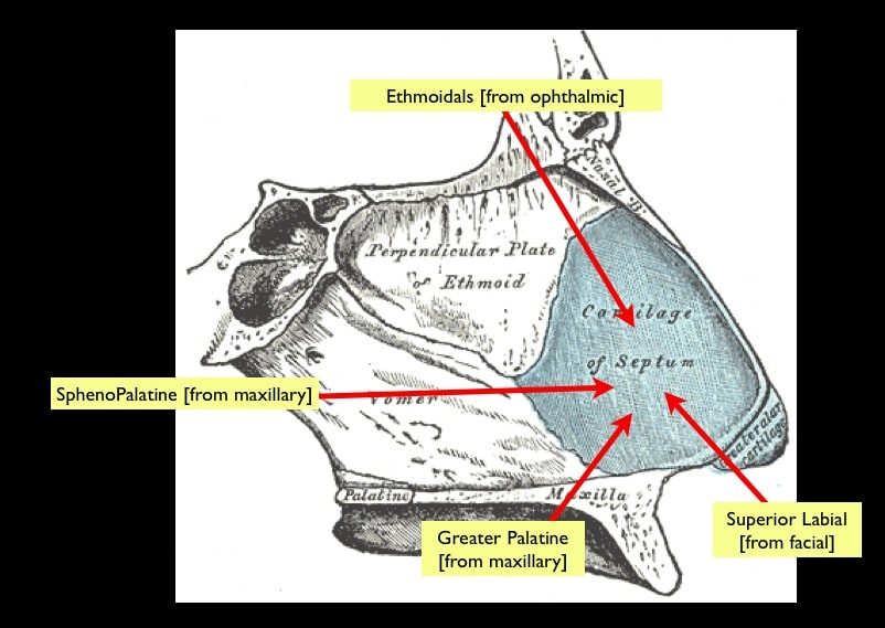
- the highly vascularised mucosa is great for drug delivery in both therapeutic and recreational forms…
Posterior epistaxis
- can be a real pain the hole, never mind being much more life-threatening. For the best talk I’ve seen, go here to EMCoreContent where there is (currently) a free video by Mel Herbert on epistaxis management that has some great stuff on posterior nose bleeds, including packing and that thing you do with putting a foley and in the posterior nasal cavity and inflating it
- endoscopically you can do cool things like this:
https://www.youtube.com/watch?v=TdSyYAvaRUs
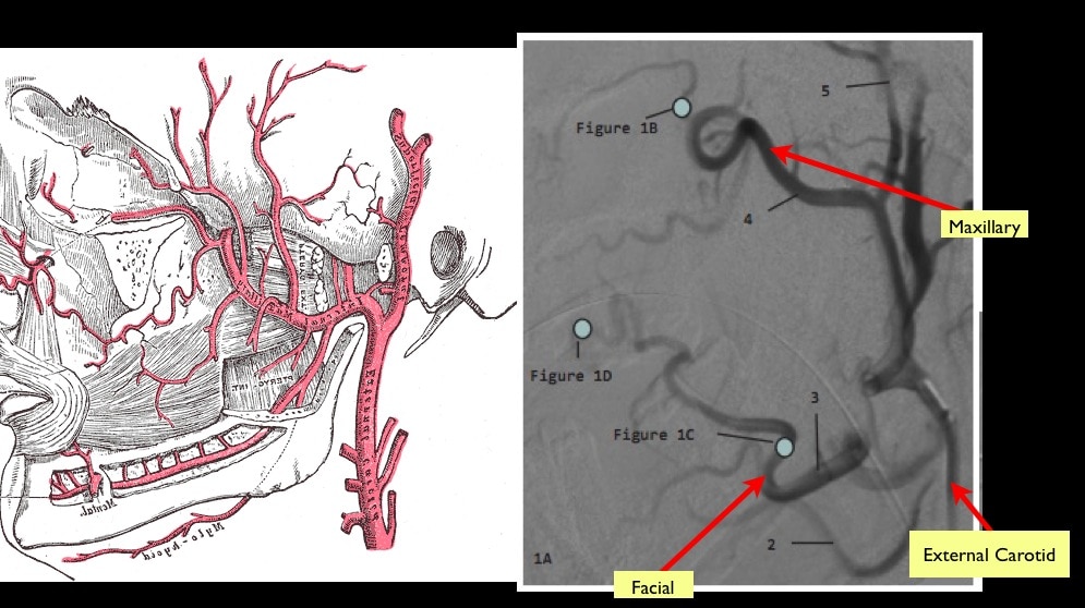
- if that fails then you can perform selective embolisation as these guys describe. I tell the students that they’ll be the generation that replaces the stethoscope with the ultrasound and the surgeon with the interventionalist…
Nasal Fractures
- Broken noses are common and thankfully it’s one thing we’ve agreed to diagnose without an x-ray
What bones do you break when you break your nose?
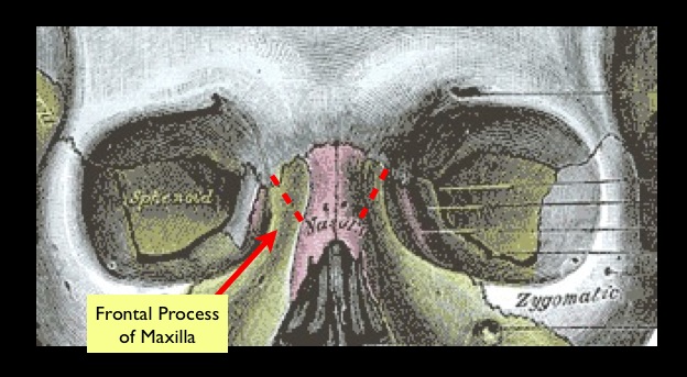
- not the nasal bones
- the fracture usually occurs at the frontal process of the maxilla
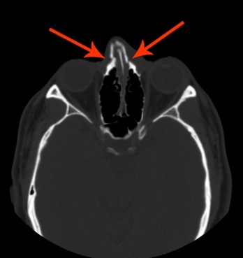
In a simple nasal fracture the thing we worry about is a nasal septal haematoma
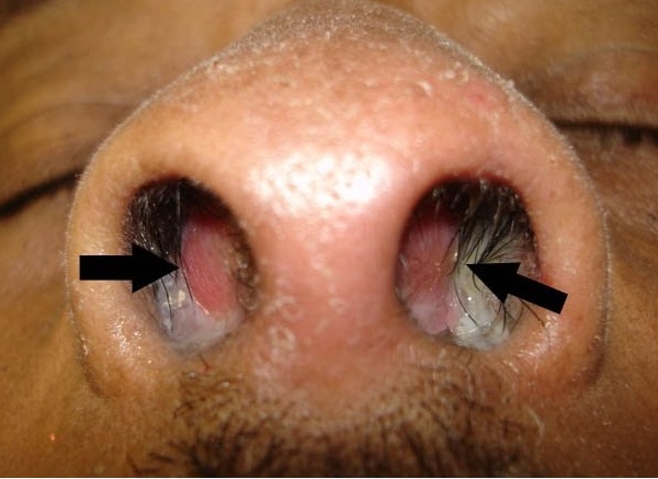
- Theory is that the submucosal bleeding on the septum strips off the perichondrium from the septal cartilage leaving the cartilage devoid of vascular supply leading to eventually necrosis of the septum
LITFL have a nice post on this with a video on manipulation of nasal fractures.

Pingback: The LITFL Review 045 - Life in the Fast Lane Medical Blog
Pingback: Näsblödning – översikt – Mind palace of an ER doc