This is a series of posts where I’m trying to transfer my anatomy lectures to a blog format, with emphasis on the clinically relevant points.
Images are mainly from the 1918 Grays, freely available and out of copyright here. Forgive the out of date nomenclature…
The AC Joint
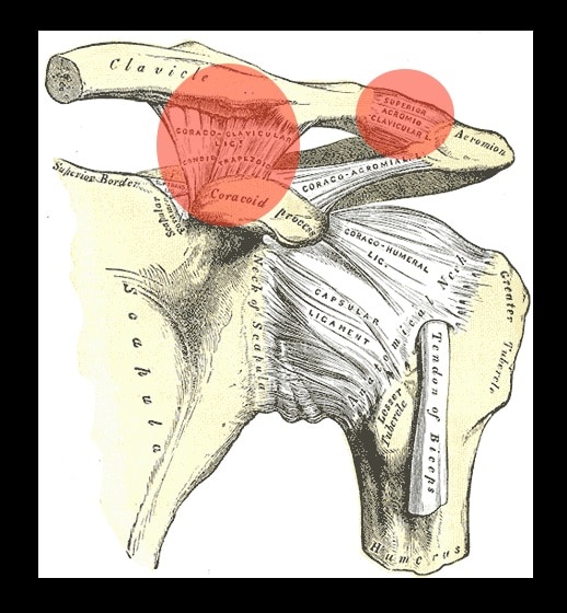
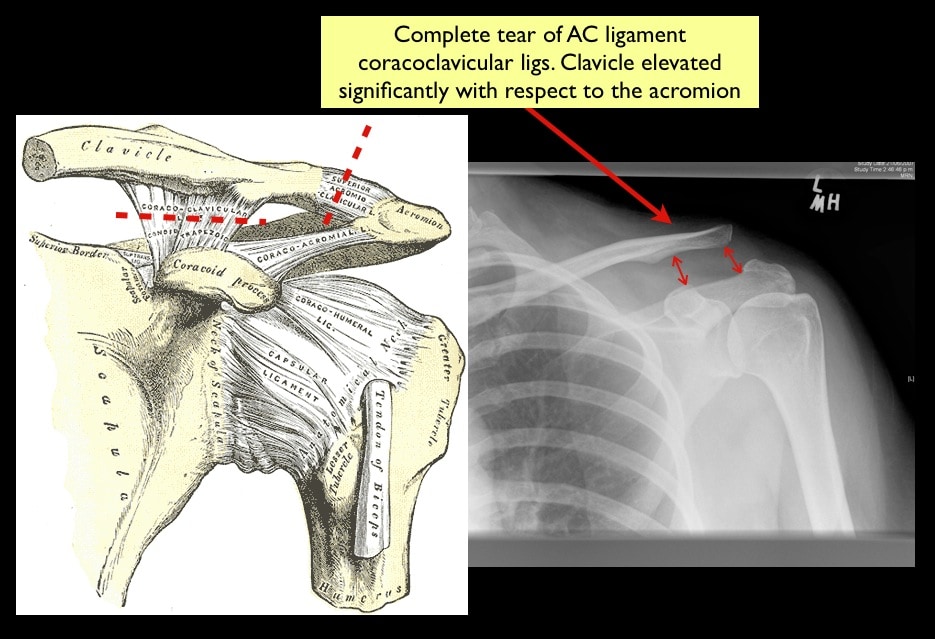
- There is no bony fit between the clavicle and acromion and so the stability of the joint relies mainly on the surrounding ligaments
- these can be divided into:
- Intrinsic/Capsular – acromioclavicular ligament
- Extrinsic – the coracoclavicular complex (trapezoid and conoid ligs)
- The AC joint itself is an odd little one. It’s synovial and lined with different cartilage than usual and has a little disc crossing the joint space. Though I can’t imagine how that information is going to be that much use to you
What goes wrong with it?
- We see a lot of AC joint injuries, normally from blows to the shoulder causing separation of clavicle and scapula
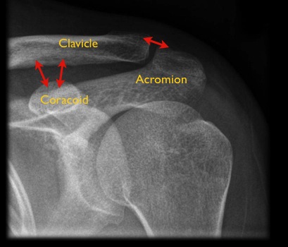
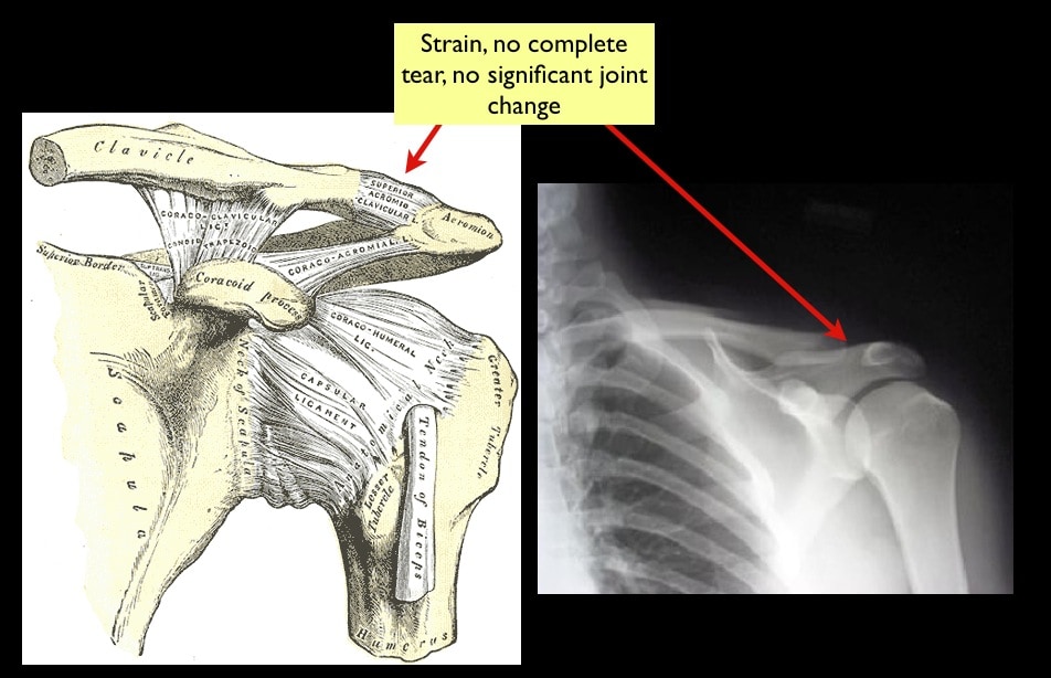
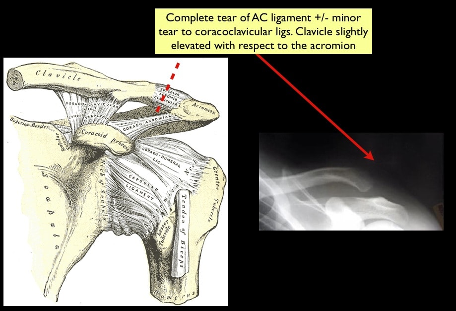
There is a grading system for injuries as follows:
The SC Joint
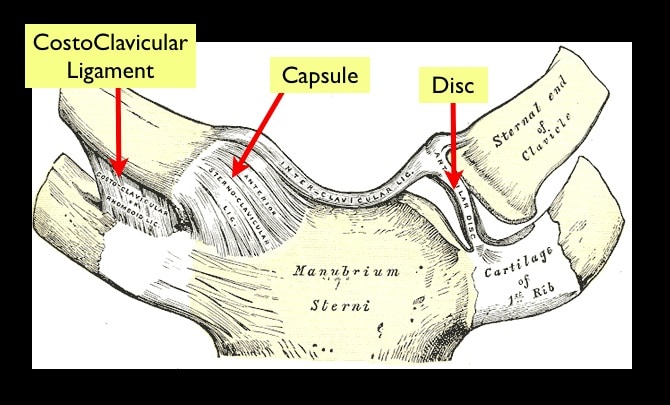
- it’s another funny little joint with a disc in it, this time the disc does the quite remarkable function of giving this join movement in 3 planes
- this is an even more inherently unstable joint than the AC joint and is actually the only bony articulation between the entire pectoral girdle and the axial skeleton
- stability comes from the strong capsule and costoclavicular ligament but also by some of the surrounding muscles (such as subclavius – anyone remember that muscle?)
On a personal note my SC joint is so unstable it subluxes anteriorly when I extend my shoulder in the abducted position. Not painful, just clunky and uncomfortable. It makes it hell to paint ceilings…
[Anterior view right SC Joint. Cropped to preserve my remaining dignity…]
https://www.youtube.com/watch?v=G_Sn-IQa5I0
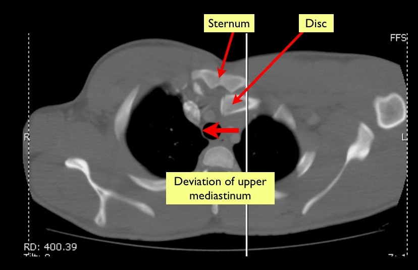
Much more seriously you can dislocate your SC joint posteriorly like this:
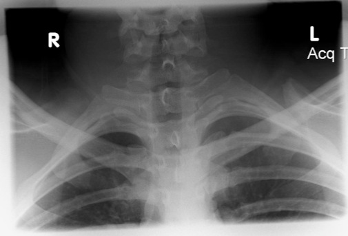
This was suspected but not really visible on the plain film or scout:
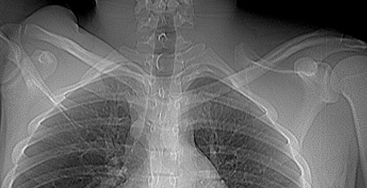
This is naturally kind of a big deal. Immediately posterior to the medial clavicle lies the subclavian vessels, never mind the trachea and oesophagus sitting medial. The patient here had some mild dysphagia but good pulses and went down the road to the big hospital for the CT and orthopaedic guys to play with.
Thank you Andy for your great anatomy notes, it puts a totally new perspective on these commonly seen problems that are so heard to learn. Just a thought, no pressure (ok just a little!) – a recording of your video lecture would be awesome… I haven’t seen any EP teaching the pearls of orthopedics this way…
Hi david
I’m not sure we have the ability to record lectures in the department, but I suppose i could do it in a vodcast format… And the lectures I give to undergrad med students are quite different from what I would do for EPs (what I’m trying to do on the blog) . I’m sure you don’t really care about the trunks and cords of the brachial plexus!
I’ll keep it on the back burner for now, or at least till i get a bit more time but it’s a good idea.
Andy