
This is a nifty little diagnosis and something that you’ll see with some regularity. In the context of sudden unexplained cardiac arrest with ROSC but no neurological response we often get a CT of the head looking for some intracranial disaster to explain what just happened.
One of the images below is a typical SAH the other is consistent with pseudo SAH, can you tell the difference? (click the radiopaedia case links to find out)
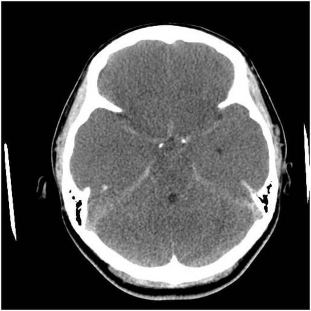
Case courtesy of Dr Chris O’Donnell, Radiopaedia.org. From the case rID: 16446
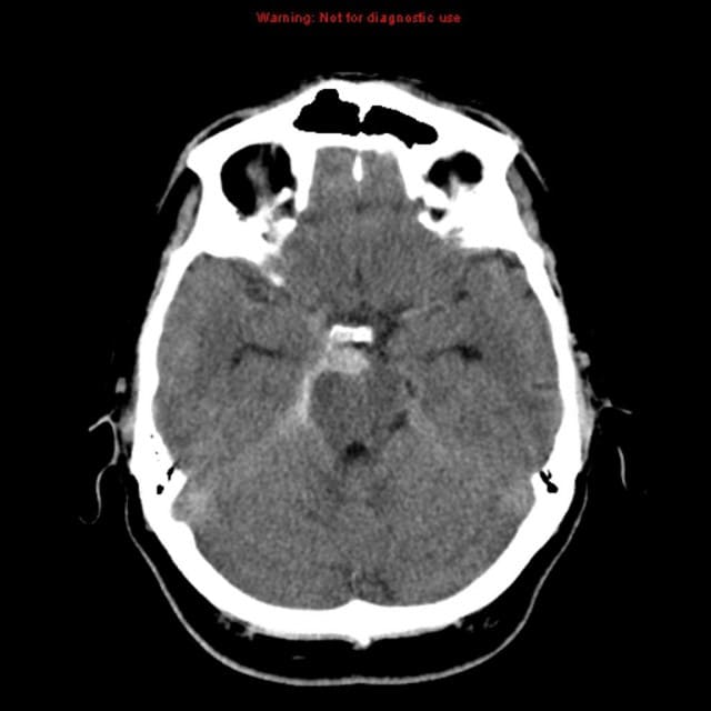
Case courtesy of Dr Saqba Farooq, Radiopaedia.org. From the case rID: 10489
I’ve seen this a couple of times now in post arrest patients and it can cause a lot of confusion as all of a sudden you’re worried about anticoagulation if you were thinking cath lab or maybe you’re tied into doing angios and what not.
Radiopaedia has a great post on it so all kudos to them but I think it’s something we in the EM world really need to know about hence the post.
In the post arrest patient (where I first encountered it) you get lots of cerebral oedema and engorgement of the superficial venous structures.
Can also occur in:
- severe meningitis
- dural venous sinus thrombosis
- bilateral large SDH
How do you tell the difference:
- ask your radiologist
- usually a lower hounsfield unit comared to true SAH
References:
- Radiopaedia
- American Journal Neuroradiology [Free full text PDF]