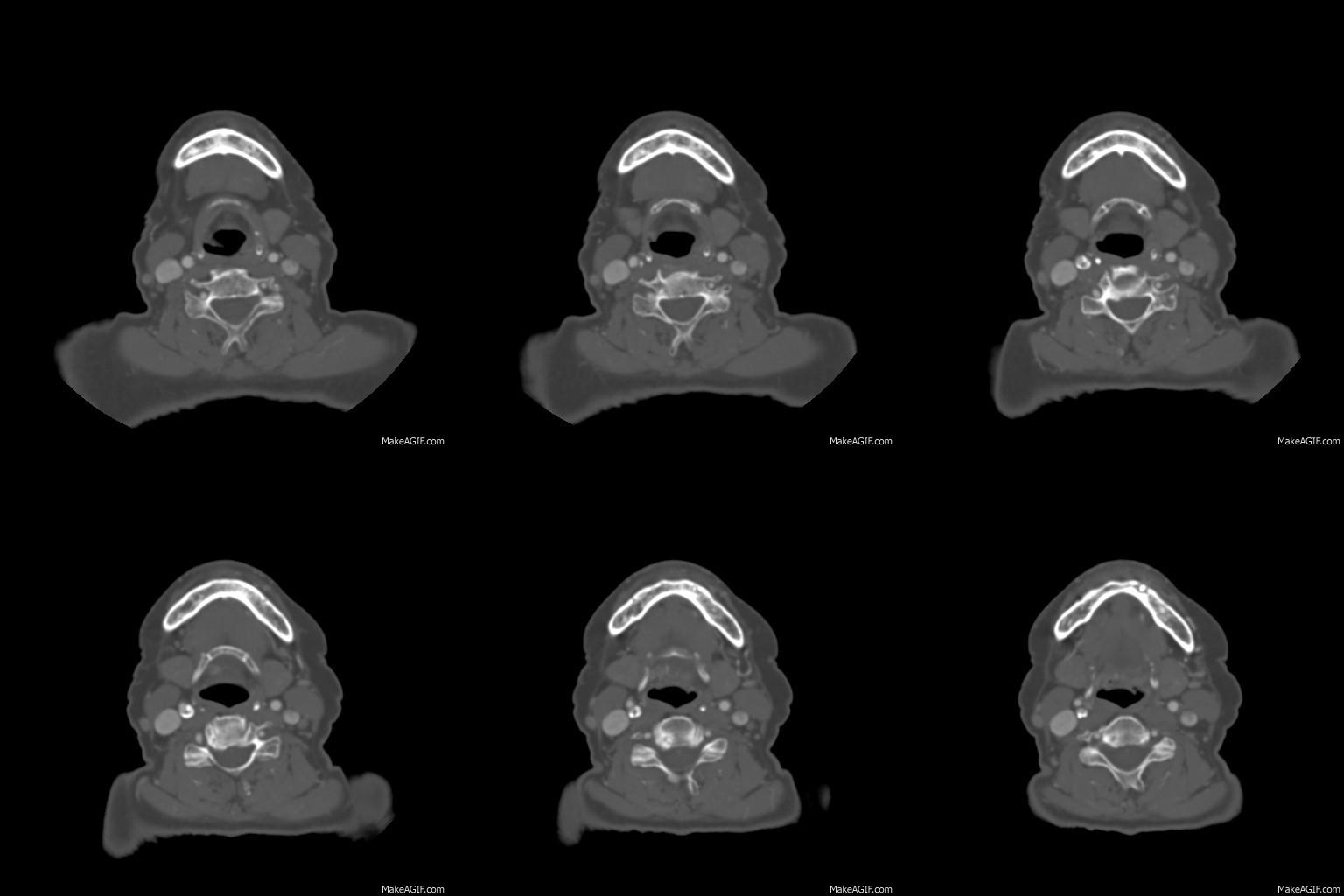
So this one is off the normal beaten track for me.
The original Scancrit post can be found here
The paper can be found here.
Here’s the rest of my thoughts on it:
This study was found by ScanCrit a while back now and generated a little bit of discussion on the twitter thingy.
Below are my notes on the study.
METHODS
- sedated pigs with lots of monitors in place
- the common carotid monitor was of course proximal to where the LMA would be so i don’t know if this affects reading seeing as we’re interested in distal flow in the ICA and not the common or the ECA
- there’s some lovely stuff on the model they used
- they performed angios on pigs with the EGDs in to see how it affected things but these were on recently dead pigs which is surely a different thing altogether
RESULTS
- flow dramatically decreased when the EGD was placed
SOME THOUGHTS
- they note that one study in anaesthesia showed reduced flow with EGD but it’s likely of little consequence there and very important in cardiac arrest
- of most importance is the fact the airway of a pig is shaped is so remarkably different that this may be entirely wrong.
- if it’s right however then reducing cerebral blood flow during a critically low flow period might be real bad news.
The podcast itself lives here:
And here’s a GIF of the CT. Sorry if the scroll is a bit slow. HT to radiology signs for the link to the MakeAGif site.


That, my friend, was an awesome post. Thanks for that!
inspired by you guys of course!
I would enjoy your presentation more if you would stop whispering and speaking in such a low, muttering tone of voice. Speak more distinctly, please. Thanks!
it seems like my mother was right all these years… fair point though – i’ll try to enunciate a little more clearly on coming episodes
Awesome post! I really got a good picture on the anatomy and posssible mechanisms behind this, IF there is something to it in humans too. And I think it is really an important question to answer, before we rush to leave the SAD and back to ETT again in cardiac arrest. My opinion is we have gained a lot more “hands-on”-time using SADs (laryngeal tubes) for prehopital providers in Norway (EMTs/paramedics with no/limited practice in intubating).
Would it be a great difference in pressure from the different types of SADs? And What about the iGel, could it be an acceptable solution? I’m really looking forward to read more on this topic!
Thank you for your post!
my experience is very limited of course and I’ve only ever used the LMA classics. there are some studies it seems on the effect of different LMAs on IJ position (for putting in lines) but there doesn’t seem to be much on its effect on flow
brilliant! The big questions to me: 1- is there anatomical impingement on the carotid? 2- does limit flow? 3- is the “oxygen-time” & cognitive offloading by putting in an LMA vs intubating in the terrible conditions of a code offset any limited flow from the balloon?
as of now I think I still prefer LMA in a code, but great discussion!
it seems those questions remain unanswered for now!
How about king LT?
Ha! Would love to say I know enough about the King LT to comment – have never used one I’m afraid.
Interestingly, in the pig study they also looked at combitubes, and the CBF was even lower!
Hi Andy,
Great post. It would be interesting to see a CT of a pig neck. Google falls short and I can’t find one. Is there a veterinary department at Trinity that could assist?
Oh, and I thought your enunciation was excellent, but maybe I have an ethnic advantage.
See you
Yeah I had a look for descriptions of comparative anatomy but there
was very little beyond some descrpitions of laryngeal anatomy rather
than vascular.
TCD has at least 3 MRIs on our building so there might be a CT in
there somewhere but unfortunately there’s layers of paperwork and
practicities to getting a pig in a scanner…
How useful would an MRI of a human with an LMA-type SAD in place be for this discussion? I am considering publishing a case report (with videos) to describe the use of the Air-Q blocker airway in 2 patients who underwent MRI under general anesthesia (whom I anesthetized). I even made 3-D reconstructions of the scans.
Hi jimmy
I’d love to see the images. I did a few pub med searches before I did the podcast looking for images and studies of LMAs and couldn’t find anything apart from studies looking at MRI compatabity of LMAs in models.
The recons would be cool too. I think it’s useful given that there’s nothing currently studied. An MRI of a pig with an LMA in would be nice for comparison. I found the angios in the pig paper really hard to interpret.
Awesome–these images are archived, so it will take me a day or so to get them out. I’ll get back to you soon!
Email is emergencymedicineireland@gmail.comlet me know
How much of a pressure wave does effective CPR produce? Would the LMA really need to produce that much lateral pressure to impede flow in a pt undergoing CPR? The length of the LMA would also play a role I Imagine. Much like the breadth of a Sphygmomanometer cuff needs to vary according to the thickness of the arm, A thin neck with little fatty tissue would potentially be more greatly effected by a larger LMA then a smaller one. I suppose this would also be limited by the protection of the carotids superiorly by the Hyoid.
In any case, a well done and interesting Post!
Good questions – i think they all remain unanswered for now at least
Pingback: IMPAIRED CAROTID ARTERY BLOOD FLOW BY SUPRAGLOTTIC AIRWAY | Dominick C. Watts